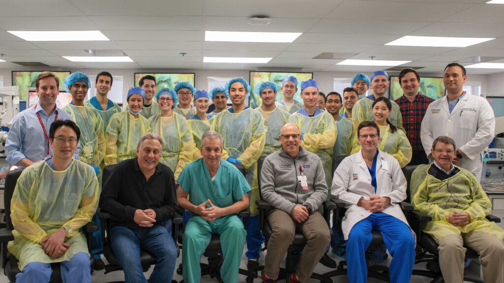
In a course taught by some of the country’s leading neurotologists and neurosurgeons, Washington University residents recently learned step-by-step the techniques to remove tumors from the skull base.
Working in Washington University’s state-of-the art multidisciplinary surgical simulation lab, residents from the departments of otolaryngology and neurosurgery practiced surgical approaches to removing an acoustic neuroma, a non-cancerous slow-growing tumor that develops on the vestibular nerve. Because neurosurgery and neurotology specialists often work closely on these surgeries, residents from both departments of otolaryngology and neurosurgery participated.
Visiting ENT surgeon Bruce Gantz, MD, prepares a demo dissection for residents as visiting neurosurgeon Paul Camarata, MD, and course leader, Michael Chicoine, MD, discuss course details.Hosted by Washington University Dec. 6-7, the event was supported by an educational grant from global medical technology company Stryker, allowing the course to invite two guest faculty members, Paul Camarata, MD, professor and chair, Department of Neurosurgery at University of Kansas, and Bruce Gantz, MD, professor and chair, Department of Otolaryngology at University of Iowa. Medical students and residents from St. Louis University and the University of Kansas Medical Center also participated.
Participating faculty from the departments of Neurosurgery and Otolaryngology at Washington University included:
- Craig Buchman, MD
- Michael Chicoine, MD
- Nedim Durakovic, MD
- Jacques A. Herzog, MD
- Albert H. Kim, MD
- Sean McEvoy, MD
- Jonathan McJunkin, MD
- Joshua Osbun, MD
- Matthew Shew, MD
- Cameron Wick, MD
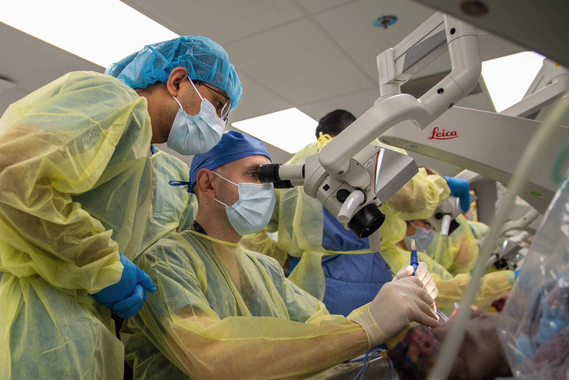
Residents gained interdisciplinary experience: neurosurgery resident Bhuvic Patel looks on as ENT resident Jay Gantz advances their skull base dissection.
Working with faculty who are national leaders in skull base surgeries, residents gained experience with three surgical approaches to the skull base. Surgery for acoustic neuromas, also called vestibular schwannoma, may involve removing all or part of the tumor.
The residents practiced the three main surgical approaches for removing an acoustic neuroma:
- The translabyrinthine approach involves making an incision behind the ear and removing bone behind the ear and some of the middle ear. This procedure is generally used for tumors larger than 3 centimeters. This approach allows the surgeon to see an important cranial nerve (the facial nerve) clearly before removing the tumor.
- The retrosigmoid/sub-occipital approach involves exposing the back of the tumor by opening the skull near the back of the head. This approach can be used for removing tumors of any size and offers the best, albeit small, possibility of preserving hearing.
- The middle fossa approach involves removing a small piece of bone above the ear canal to access and remove small tumors confined to the internal auditory canal, the narrow passageway from the brain to the inner ear. This approach is recommended for patients with smaller tumors and good hearing in the affected ear.
 “This year’s lateral skull base course set a new high bar for excellence in surgical training because of the generous support of Stryker, the great lectures and teaching by our two outstanding visiting faculty, and the participation of many faculty from ENT and neurosurgery from Washington University,” said course leader and Professor of Neurosurgery Dr. Chicoine.
Guest lecturer Dr. Gantz commented on the interdisciplinary program: “This was one of the best dissection lab experiences that I have participated in.”
For second year ENT resident, Ethan Craig, MD, the lateral skull base course was a valuable experience. “The opportunity to walk through a cadaveric dissection of complicated anatomy under the guidance of world class surgeons was amazing,” he said. “I especially appreciated the input from multiple disciplines and a unique and shared surgical space.”