For more than 30 years, Mark Warchol, PhD, been interested in the development and repair of sensory hair cells of the auditory and vestibular systems, crucial to hearing and balance. That search has led the Warchol lab to identify an unlikely contributor to these processes – scavenger cells known to clean up debris from cell death, called the macrophage.
Sensory hair cells of the inner ear are critical because they convert energy from movement and sound into neural signals transmitted by the vestibular and auditory neurons. Once hair cells are damaged, they cannot regenerate to replace dead cells, so hearing and balance health is affected for life.
Enter the scavengers to remove the ‘corpses’
Macrophages are scavenger cells that are well known for their ability to clean up the debris that results from cell death or damage. Macrophages of the inner ear are derived from white blood cell precursors and enter the ear early in its embryonic development.
As one component of our innate immune system–a collection of cells and tissues that protect us against foreign pathogens–these cells may play a more critical role for the inner ear than simply clearing debris.
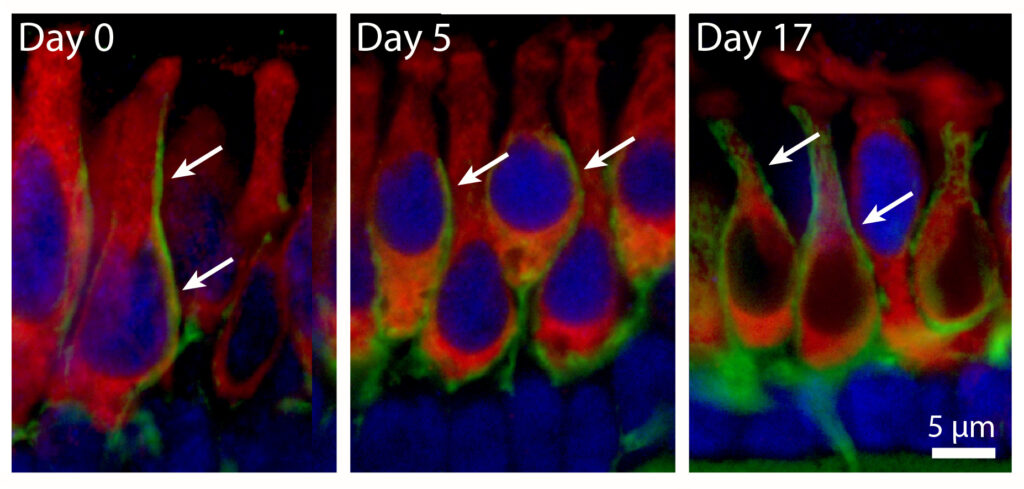
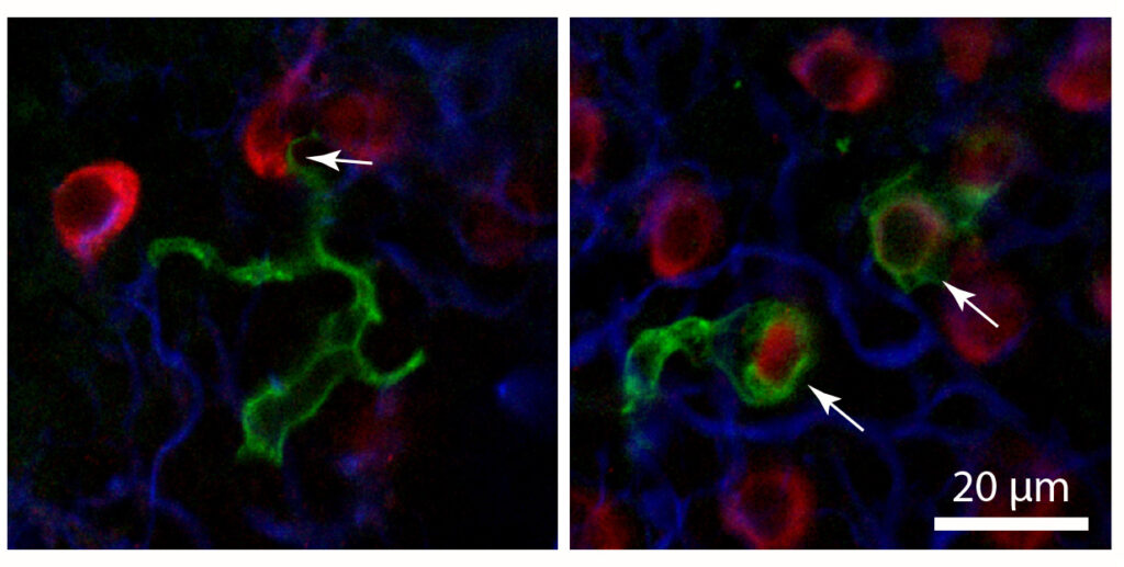
According to Warchol, one function of macrophages in the injured ear is to remove the ‘corpses’ of dead cells, a process that helps preserve the structure of the inner ear after injury.
“I think we published the first images of macrophages actually engulfing dying hair cells,” he said. “We are currently trying to figure out how macrophages distinguish between dying cells and healthy cells.”
Current studies in the lab are aimed at answering several questions:
- What chemical signals attract macrophages to sites of injury in the inner ear?
- Once they arrive, how do macrophages distinguish between dying cells and healthy cells?
- Do macrophages play a role in the embryonic development of the inner ear?
‘Sculptors’ to the rescue?
In a separate project, the Warchol lab is studying the role of macrophage in the embryonic ear. The number of macrophages in the developing cochlea is about three times greater than in the mature ear, and those numbers peak at times when the cochlea is undergoing significant changes in its structure and innervation.

“The cochlea is a very precisely-patterned structure, and we hypothesize that macrophages may serve as ‘sculptors’ during cochlear development, by removing unnecessary cells,” said Warchol.
Additional efforts are aimed at understanding how the anti-cancer drug cisplatin damages the sensory cells of the inner ear. Cisplatin is among the most widely used antitumor drugs, but one that causes permanent hearing loss in significant numbers of patients who undergo chemotherapy. In collaboration with Lavinia Sheets, PhD, the lab wants to know how inflammation influences the extent of cisplatin ototoxicity.
“I have always taken a comparative approach to the study of the inner ear,” says Warchol. “Our current projects utilize zebrafish, chickens and mice.”
“The collegial environment of our department has been very helpful in extending these studies to other species,” said Warchol. “We have collaborated with Dr. Keiko Hirose, Dr. Kevin Ohlemiller, and Dr. Mark Rutherford on studies of the mouse inner ear, and with Dr. Sheets on several projects that involve zebrafish.”
