Washington University otolaryngology residents Amit Walia, MD, and David Lee, MD, received research grants from the American Hearing Research Foundation (AHRF). The awards are $1,000 each and will provide supplies and other expenses related to their research projects.
Title: Optical Coherence Tomography of Inner Ear Structures
PI: David Lee, MD
Mentor: Nedim Durakovic, MD
Hearing instability disorders, like Meniere’s disease and sudden sensorineural hearing loss (SSNHL), are poorly understood and inadequately treated. Steroid therapy has been the standard of care for decades, but results have been inconsistent. Improvements in patient outcomes have been challenged by an inability to detect inner ear pathology, where hearing instability disorders are thought to occur. Otolaryngologists are currently forced to rely on patient history and audiograms to guide clinical decisions. This makes diagnosis ambiguous and treatment responses difficult to evaluate.
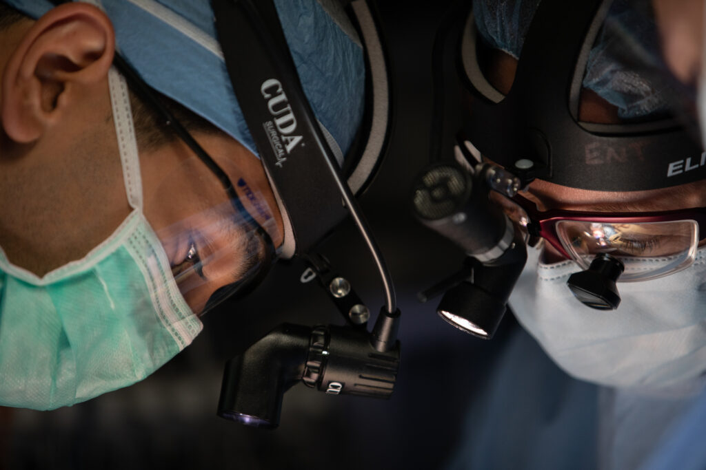
Imaging is an attractive method for monitoring disorders of hearing instability because it is non-invasive and provides objective data. However, current imaging techniques, like CT and MRI, are unable to adequately view the small structures within the inner ear. Optical coherence tomography (OCT) is a newer imaging modality that might measure structures of the inner ear with sufficient resolution to diagnose and monitor diseases in these tissues. OCT measures the reflectance profile of infrared light emitted from a fiber optic probe to enable high-resolution images in real time. Since OCT can detect and quantify changes at the microstructure level, it may be able to detect small changes within the cochlea that likely occur in the setting of Meniere’s disease or SSNHL. This project seeks to use this new imaging modality to view structures of the inner ear and improve diagnosis and monitoring of hearing instability disorders.
Title: Using electrocochleography to build a prediction model for speech-perception performance in noise after cochlear implantation
PI: Amit Walia, MD
Mentors: Craig Buchman, MD, Jacques Herzog, MD, Matthew Shew, MD, and Amanda Ortmann, PhD
Cochlear implantation (CI) has become an effective treatment option over the last three decades for those with severe-to-profound hearing loss. Although many patients do well after implantation, performance as measured by speech perception outcomes is highly variable and remains largely unexplained. Recognizing factors that may limit speech perception performance after implantation would be useful in counseling patients preoperatively, tailoring a patient-specific aural rehabilitation pathway, and improving surgical approaches and customizing electrode designs.
Electrocochleography measures the electrical responses of the cochlea and auditory nerve to sound stimuli to assess hearing function. This test has been used to detect and monitor inner ear pressure (endolymphatic hydrops) and to monitor auditory function during surgery. The goal of this project is to utilize electrocochleography to measure acoustically evoked electrical potentials from the cochlea and quantify residual cochlear function with the goal of predicting performance in quiet and background noise prior to electrode array insertion.