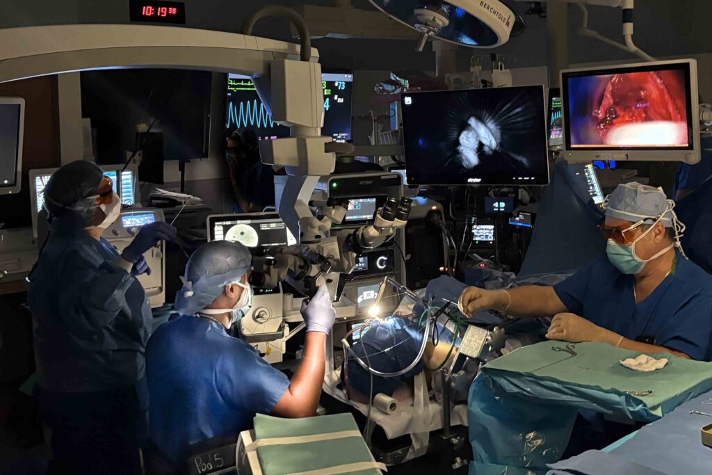
WashU Medicine voice and airway surgeon Bharat Panuganti, MD, is adapting a fluorescent imaging technology known as confocal laser endomicroscopy (CLE) to better view cellular detail and optimize laryngeal cancer removal.

One of the consistent challenges during surgical removal of any cancer is delineating tumor from surrounding normal tissue. Conservative approaches suggest that to error on the side of excess tissue removal reduces the risk of recurrence, but this practice also increases the likelihood of cosmetic or functional deficits.
“Laryngeal cancers represent a challenging surgical entity — the larynx is a complex three-dimensional organ where even small amounts of tissue loss can manifest with outsized functional consequences,” said Panuganti. “This complicates the prospect of accurate margin assessment and function preservation during larynx tumor resections.”
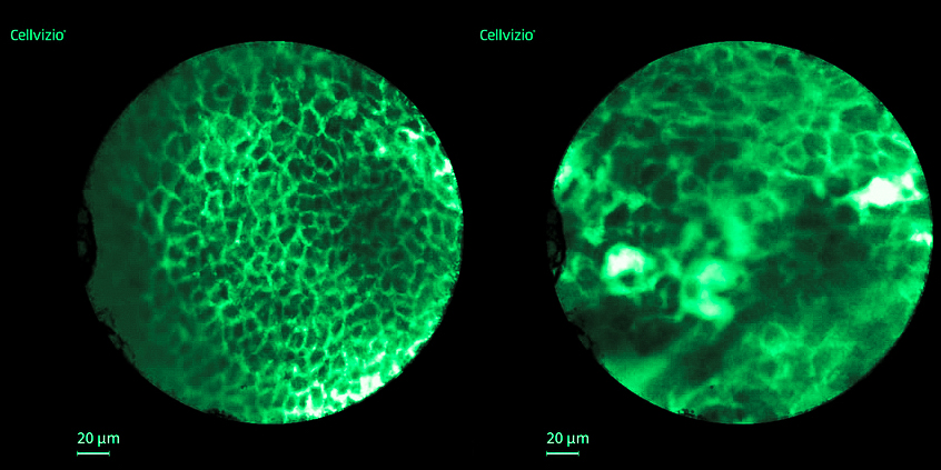
He further explained that fine details like the patterns of cellular organization are poorly seen even with high fidelity surgical endoscopes, which makes differentiating normal tissue from dysplastic cancer tissue difficult. CLE shows surgeons a thin optical slice of tissue, enabling a much better appreciation of cell size, shape and organization.
Now, I don’t have to make assumptions about appropriate cancer margins, but can actually decide with the benefit of in vivo microscopic optical biopsies.
Bharat Panuganti, MD
“We use 10% fluorescein as an intravenous dye to highlight cellular morphology in real time and infer the probability of cancer or pre-cancer in regions of concern,” he said. “Now, I don’t have to make assumptions about appropriate cancer margins, but can actually decide with the benefit of in vivo microscopic optical biopsies.”
Panuganti stated that CLE represents just the latest tool in the arsenal of intraoperative imaging for larynx cancer at WashU Medicine. It has now been utilized in ten laryngeal cancer cases since its arrival six weeks ago, and the team is actively exploring expanding CLE usage to other head and neck mucosal subsites.
“We are constantly learning more about the benefits of imaging on a microscopic scale,” he said. “This approach allows us to identify concerning microscopic changes in regions of otherwise grossly normal mucosa. This provides a unique opportunity to hone the practice of ‘precision surgery’ based less on presumption and more on a highly detailed understanding of microscopic disease.”
To learn more about laryngeal cancer or confocal laser endomicroscopy, contact Bharat Panuganti, MD, at bharatp@wustl.edu.