Panuganti Lab
Principal Investigator:
Bharat Panuganti, MD, FACS, assistant professor of Otolaryngology–Head and Neck Surgery
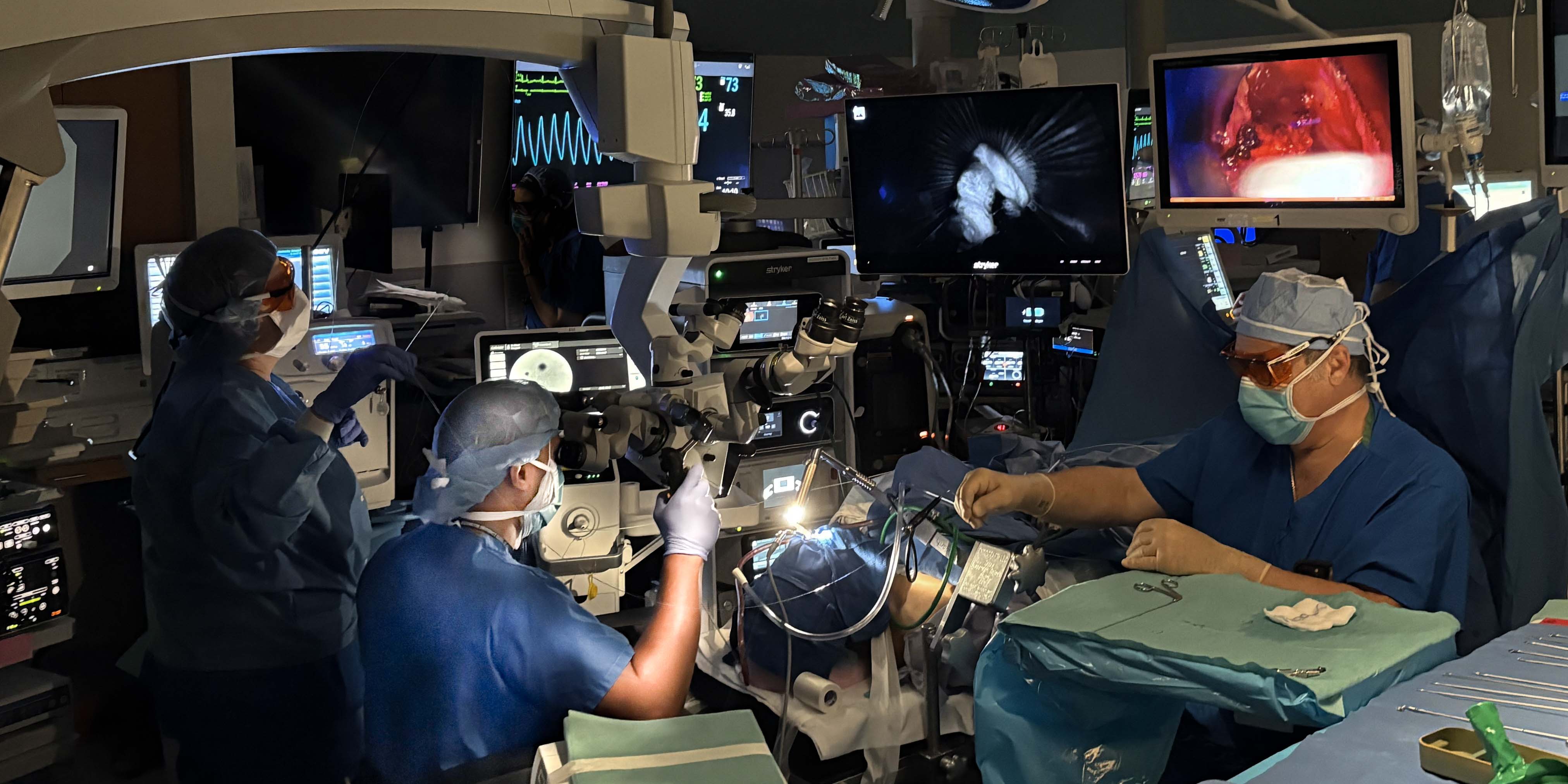
Our research centers on improving surgical care for laryngeal cancer and complex airway disease through the development of advanced intraoperative imaging and innovative surgical techniques.

Our goal is to provide surgeons better visualization of tumor margins in real time during transoral laser and robotic surgery, where conventional pathology is often limited. We hope to show that optical imaging technologies like fluorescence-guided surgery and confocal laser endomicroscopy will enable more precise, tissue-sparing resections.
I also lead interdisciplinary efforts applying artificial intelligence and multimodal image integration to create dynamic intraoperative tumor maps.
In parallel, my research includes translational studies in airway therapeutics and molecular characterization of laryngeal disease, with the overarching goal of improving oncologic outcomes while preserving voice and airway function.
Research Projects
Combination optical imaging to guide transoral laser microsurgery in larynx cancer
Funding: NIH R21 (Award #1R21CA292196-01)- $374,880 (Co-PI: Jason Warram, PhD)
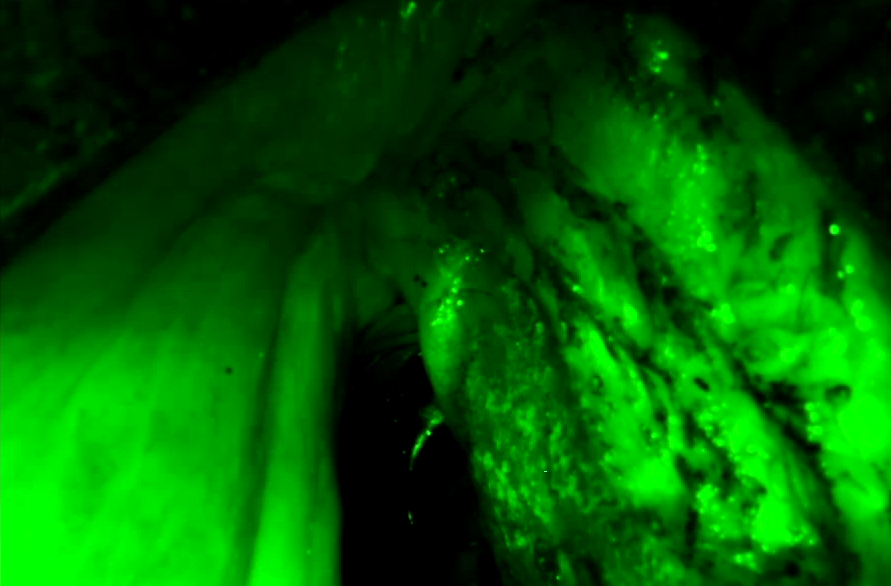
Overview: To establish metrics for a Phase II clinical trial evaluating combination intraoperative targeted fluorescence guided surgery (FGS) and probe-based confocal laser endomicroscopy to guide resection margins and post-resection margins in patients with laryngeal cancer. To elucidate incremental efficacy of detecting clinically occult disease, originating from peripheral mucosa and deep submucosae, using combination optical imaging.
Assessment of safety and residence time of sustained-release triamcinolone formulation in polylactic-co-glycolic acid (PLGA) microspheres in the rat subglottic submucosal compartment
Funding: American Laryngological Association Seymour Cohen Research Grant- $15,000
Overview: To assess biocompatibility and residence time of a unique long-acting steroid formulation embedded in PLGA microspheres insinuated in the submucosal compartment of the rat trachea, in preparation for a phase I human trial.
Integrative Sparse Feature Matching, Spatial Analysis, and AI-Based Tissue Characterization for Comprehensive Multi-Modal Tumor Mapping in Laryngeal Cancer Management
Funding: Washington University in St. Louis, ICTS Precision Health Grant- $25,000 (Co-PI: Nathan Jacobs, PhD)
Overview: Collaborative endeavor to develop a computation platform that can leverage machine learning techniques to extract and correlate key features from multiple imaging modalities in real time, despite inherent differences in resolution and perspective, to generate an integrated, dynamic intraoperative tumor map based on information gleaned from a spectrum of optical imaging sources.
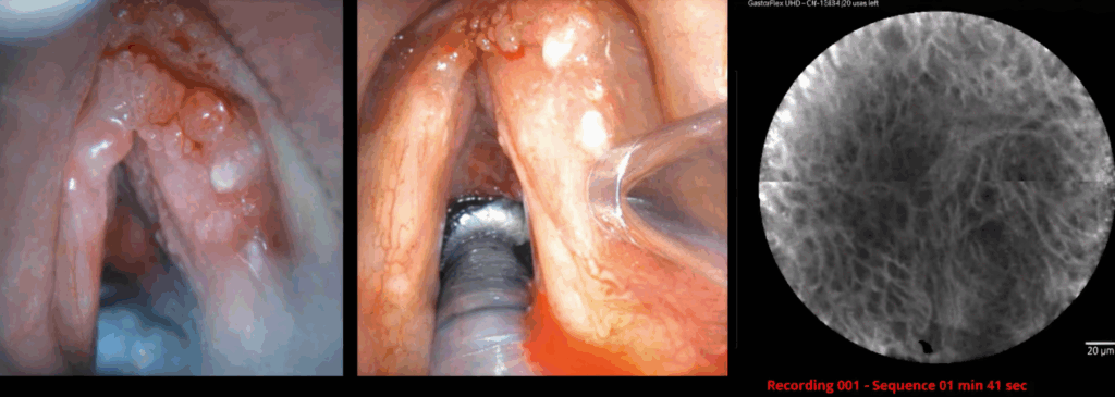
Fluorescein-Enhanced Confocal Endomicroscopy (CLE) for Real-Time Tumor Mapping and Margin Assessment in Head & Neck and Brain Cancer Surgery
Funding: Barnes Jewish Hospital Foundation – $230,000 (Co-PI: Dimitrios Mathios, MD)
Overview: Interdepartmental collaboration exploring incremental value of CLE with fluorescein during head and neck mucosal, salivary gland, and brain tumor resections, assessed via relative impact on local recurrence rates and functional outcomes.
Publications
Visit Dr. Panuganti’s WashU Profile to see a complete list of publications and collaborations»