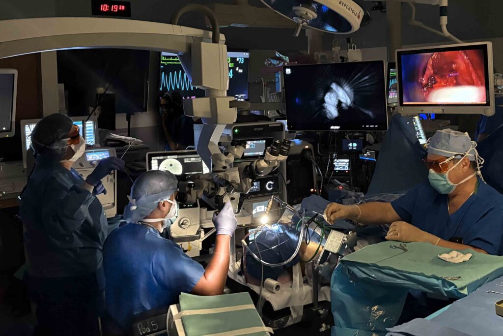
Welcome to Innovations in Otolaryngology, a series of feature stories to highlight the pioneering advances in patient care being researched and implemented by the Department of Otolaryngology – Head and Neck Surgery at WashU Medicine.

In today’s feature, laryngeal surgeon and Assistant Professor of Otolaryngology Bharat Panuganti, MD, shares recent efforts to improve intraoperative imaging of laryngeal cancer to guide precision surgical removal.
Intraoperative imaging challenged by position and complex nature of the larynx
Intraoperative visualization of the larynx has long suffered due to its position deep in the neck. This, compounded by a uniquely complex architecture and multiple tissue layers, has resulted in surgical tumor resection that is often overly aggressive or leaves tumor behind. Current positive margin rates following laryngeal cancer resection average between 20 and 50 percent, an unacceptable level according to Panuganti.
Even with vastly improved fidelity of modern endoscopic imaging devices, the challenge of distinguishing microscopic disease during laryngeal cancer resections is significant. The presence of infection, edema and radiation-induced fibrosis only further complicates the task of discerning tumor from normal tissue.

Panuganti’s goal has been the development an optical imaging protocol that would define the tumor footprint and allow precision surgery. Further, he wanted the protocol to be able to take advantage of readily available fluorescent dyes that could be administered intra-operatively, using resources readily available to most surgeons.
Two different approaches: label the tumor or show it to be different
Panuganti’s first approach has been to use a fluorescent dye called indocyanine green or ICG. Injected into the vasculature, ICG demonstrates rapid distribution and leaks from the vessels into normal and tumor interstitial spaces. The dye however dissipates quickly – within seconds – from normal tissue, but resides in tumor longer. A pilot study of twenty-seven patients undergoing transoral laser microsurgery with indocyanine green imaging suggests that fluorescence is significantly greater both in gross and occult tumor, compared to background fluorescence of normal tissue.
Another method, called confocal laser endomicroscopy or CLE, takes advantage of higher-powered microscopy and fluorescent dyes that reveal the cellular and vessel architecture of both normal and tumor tissues. Fluorescein, a common fluorescent dye, is injected intravenously and a probe placed on the surface of the laryngeal tissue reveals cell and vessel architecture to a depth of 70-130 micrometers.

Using this technique, normal cell morphology can be differentiated from tumor on a microscopic scale. The probe can be used to easily scan the entire mucosa, reducing the likelihood of missing even small regions of tumor. A recent study by Panuganti has shown CLE can improve tumor identification by more than 50 percent, compared to the use of standard white light imaging alone.
On the horizon
While the techniques described above are favored for their ready availability and intra-operative utility, Panuganti acknowledges there are other methods that might more directly label laryngeal cancer.
Panatimumab-IRDye800 is a tumor-specific antibody conjugated to a fluorescent marker. It has been used in a few cases by Panuganti and shows promise for precisely identifying the tumor footprint. Its use is constrained by issues of availability, cost and patient access – the marker must be administered to patients 24-48 hours prior to surgery. Future clinical trials assessing the long-term oncologic and functional impact of combined intraoperative optical imaging in the care of laryngeal cancer are awaiting NIH funding.
To learn more about these studies, please contact Bharat Panuganti, MD, at bharatp@wustl.edu.