Sheets Lab
Principal investigator:
Lavinia Sheets, PhD, Assistant Professor, Otolaryngology – Head & Neck Surgery
Overview
Hair cells are the sensory receptors of sound, motion, and spatial orientation. Overexposure to sound initiates a series of molecular events in hair cells that lead to various pathologies. Our research interests are to understand how specific pathological changes occur in hair cells exposed to noise by defining the dynamic cellular processes that lead to hair-cell synapse loss and hair-cell death. We investigate these questions using zebrafish as a model for human hearing and deafness.
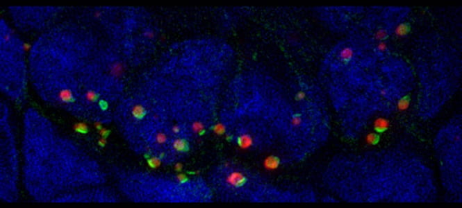
Super-resolution microscopy image of ribbon synapses—the specialized synapses found in hair cells. Presynaptic ribbons are shown red, postsynaptic densities are shown green.
Our research — in depth
Sensory hair cells—the sensory receptors of the auditory system—must convey a wide range of sound intensities (from a whisper to a thunderclap) and do so both reliably and inexhaustibly. This is accomplished by exquisitely sensitive synaptic connections between hair cells and nerve fibers that carry sensory information to the brain. The sensitivity of hair-cell synaptic connections comes at a price—they are vulnerable to damage when exposed to intense noise. Recent research indicates prolonged exposure to moderately loud noise, such as a rock concert or a stadium football game, damages hair-cell synaptic connections, resulting in hair-cell synapse loss and permanent deficits in auditory acuity.
Zebrafish are a well-established model for human hearing and deafness and provide an advantageous system to investigate mechanisms of noise-induced hair-cell damage. The zebrafish lateral-line organ—a sensory organ used to detect the movement of water—contains superficially localized hair cells that are relatively easy to manipulate pharmacologically or genetically, and are optically accessible in whole larval fish. We can thereby manipulate cellular pathways in hair cells and examine progressive hair-cell damage in a live, intact preparation. In addition, the Sheets lab takes advantage of the zebrafish’s ability to regenerate complex tissues following damage, including hair-cells and their innervating nerve fibers.
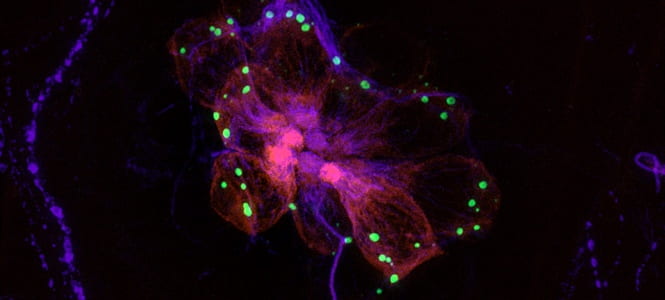
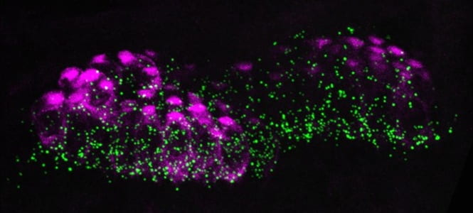
A lateral view of a larval zebrafish lateral-line organ. Radially-organized hair cells (magenta) form synaptic connections (green) with innervating nerves (purple).
Research projects
- Defining the cellular pathways that bring about hair-cell synapse loss and hair cell death following damaging noise exposures.
- Identifying biological pathways that promote nerve regeneration and hair-cell reinnervation with the goal of providing information toward clinical regenerative therapies.
- Identifying novel protectants against cisplatin-induced neurotoxicity with the goal of protecting auditory nerve fibers from damage during cisplatin-based chemotherapy.
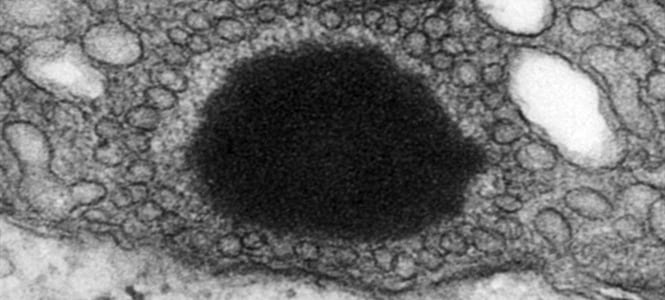
Transmission electron micrograph of a zebrafish hair-cell ribbon synapse. The electron-dense ribbon appears black and is surrounded by tethered synaptic vesicles. This specialized synapse plays an important role in transmitting sensory information.
Lab Team
- Kyle Newton, PhD, Postdoctoral Associate
- Melanie Holmgren, Research Technician
- Allison Saettele, Research Technician
- Angie Schrader, Research Specialist
Funding
NIH Research Project Grant (R01DC016066)
Roles of the Synapse in Hair-Cell Pathology • 07/01/17-06/30/22
The goal of this research is to understand how pathological changes at the hair-cell synapse stemming from excessive noise exposure ultimately contribute to sensorineural hearing loss.
Action on Hearing Loss International Grant
Identifying Novel Therapies to Promote Nerve Regeneration and Hair-Cell Nerve Reinnervation • 03/01/17 – 3/01/20
The goal of this project is to identify compounds that may promote cochlear nerve regeneration and hair-cell reinnervation by performing a large-scale drug screen for modulators of lateral-line organ regeneration in zebrafish.
Children’s Discovery Institute
Prevention of Sensory Pathology following Cisplatin Chemotherapy • 07/01/18 – 6/30/21
The goal of this project is to identify novel protectants against cisplatin-induced neurotoxicity by performing a large-scale drug screen in zebrafish. This screen will not only provide information toward pharmacological interventions that may protect auditory nerve fibers, but will also identify compounds that may protect against general cisplatin-induced sensory neuropathy.
Select Publications
Sheets L, He XJ, Olt J, Schreck M, Petralia RS, Wang YX, Zhang Q, Beirl A, Nicolson T, Marcotti W, Trapani JG, Kindt KS. Journal of Neuroscience. 2017 June 28, 37 (26) 6299-6313; DOI: https://doi.org/10.1523/JNEUROSCI.2878-16.2017
Sebe JY, Cho S, Sheets L, Rutherford MA, von Gersdorff H, Raible DW. Journal of Neuroscience. 2017 Jun 21;37(25):6162-6175. doi: 10.1523/JNEUROSCI.3644-16.2017.
Excessive activation of ionotropic glutamate receptors induces apoptotic hair-cell death independent of afferent and efferent innervation. Sheets L. Sci Rep. 2017 Jan 23. 7:41102. doi: 10.1038/srep41102.
Synaptic ribbons require ribeye for electron density, proper synaptic localization, and recruitment of calcium channels. Lv C, Stewart WJ, Akanyeti O, Frederick C, Zhu J, Santos-Sacchi J, Sheets L, Liao JC, Zenisek D. Cell Rep. 2016 Jun 21. 15(12):2784–95. doi: 10.1016/j.celrep.2016.05.045.
Characterization of Ribeye subunits in zebrafish hair cells reveals that exogenous Ribeye B-domain and CtBP1 localize to the basal ends of synaptic ribbons. Sheets L, Hagen MW, Nicolson T. PLoS One. 2014 Sep 10. 9(9):e107256. doi: 10.1371/journal.pone.0107256.
Presynaptic CaV1.3 channels regulate synaptic ribbon size and are required for synaptic maintenance in sensory hair cells. Sheets L, Kindt KS, Nicolson T. Journal of Neuroscience. 2012 Nov 28. 32(48):17211–17224
Ribeye is required for presynaptic CaV1.3a channel localization and afferent innervation of sensory hair cells. Sheets L, Trapani JG, Mo W, Obholzer N, Nicolson T. Development. 2011 Apr. 138(7):1309–19.
A full list of publications can be found at https://www.ncbi.nlm.nih.gov/pubmed/?term=lavinia+sheets
Contact Us
Mailing Address
Dept. Otolaryngology, Campus Box 8115
Washington Univ. School of Medicine
660 S. Euclid Ave.
St. Louis, MO 63110
sheetsl@wustl.edu
Physical Address
CID Building
Room 2243 (lab), 2258 (office)
4560 Clayton Ave.
St. Louis, MO 63110
Confocal image of a larval zebrafish hearing organ.