The new state-of-the-art surgical simulation lab on the medical school campus of Washington University in St. Louis provides surgical trainees access to a risk-free environment in which to practice surgical procedures.
But it’s not just for surgery.
Audiology and Deaf Education students in WashU’s Program in Audiology and Communication Sciences (PACS) are taking advantage of the lab to review anatomy of the head and neck as part of their first-year course, Anatomy and Physiology of Speech and Hearing.
The collection of anatomical models and human specimens provides the students a unique hands-on approach to explore the structures they have only viewed in two dimensions as part of their classroom lectures.
Course instructor Brian Faddis, PhD, also manages the simulation lab. He has taught the course for almost 30 years and was eager to take advantage of the new facility.
“For years I lugged models and specimens over to the PACS classrooms in the CID Building,” he said. “Bringing the students to the laboratory provides them a more immersive experience by opening a wider array of equipment and other resources they can use.”
During the semester-long course, students will visit the lab three times, to review:
- head and neck anatomy, including bones and muscles of the region, as well as structures of the outer, middle and inner ear;
- the human brain; and
- airway structures related to speech and respiration.
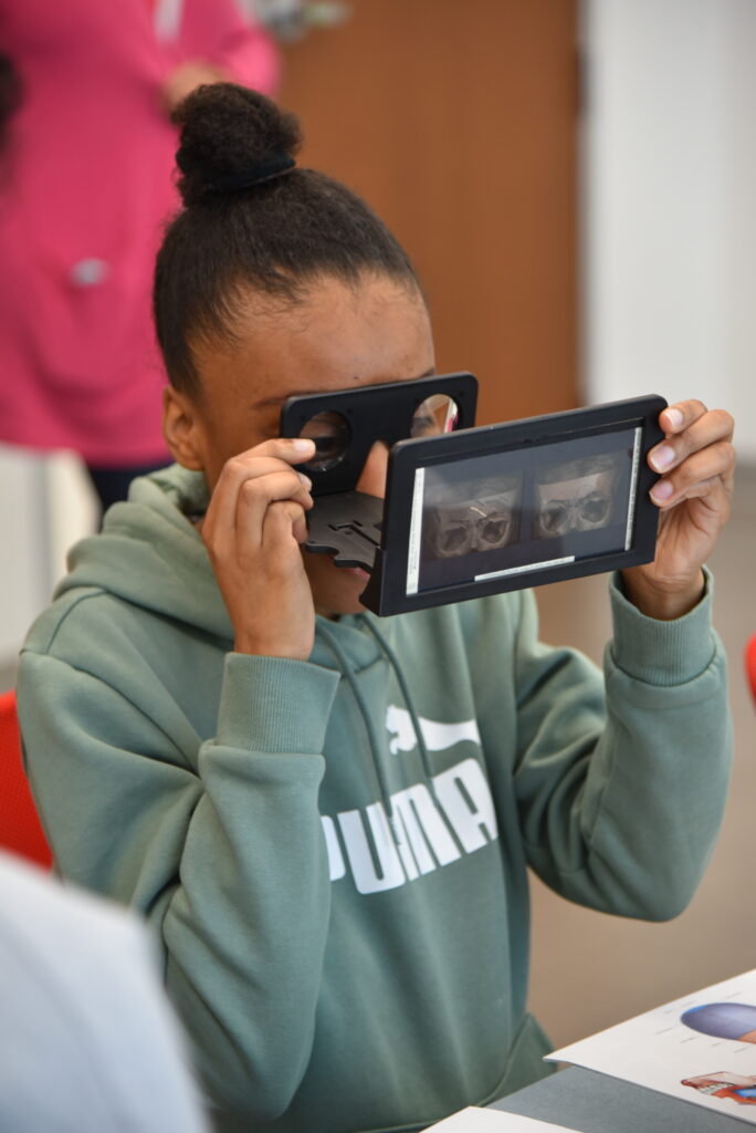
Students also enjoy viewing a unique collection of stereoscopic slides of anatomic preparations. The collection was obtained by Max Goldstein, MD, founder of Central Institute for the Deaf, when he studied in Vienna in the early 1890s.
Audiology student Stephanie Bertino was surprised by what she saw.
“We always hear about these structures being so small they can fit on a dime, but getting to see the ossicles and cochlea with our own eyes it was hard to believe.” she said. “It makes the whole process of hearing even more fascinating because these tiny bones are what we rely on every day!”
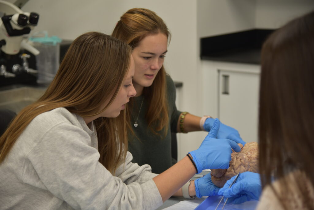
Undergraduate student Gabriela Rojo loved the brain lab.
“It was so helpful to see and interact with the anatomy,” she said. “We were told to expect a few differences between the brains but seeing it up close in person was so cool! It was an amazing experience to be able to touch the brains and recognize the structures we have been learning.”
Though the lab was specifically designed for use by trainees in Otolaryngology, Neurosurgery and Ophthalmology, Faddis would like to see more campus groups take advantage of the facility. To learn more, visit the lab webpage.