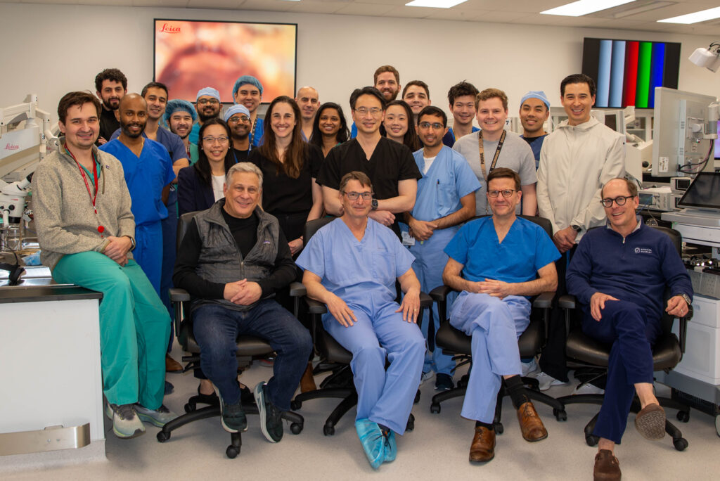
Mentored by leading neurotologists and neurosurgeons, WashU Medicine residents recently learned techniques to remove tumors from the lateral skull base.
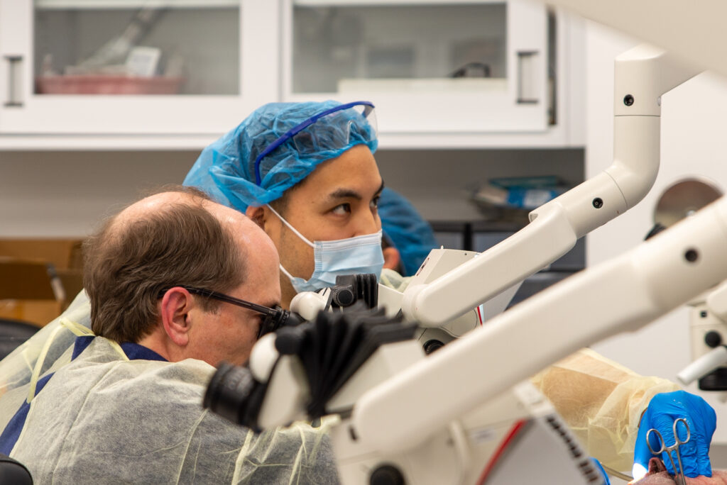
Working in the new, state-of-the art Multidisciplinary Surgical Simulation Lab in the Farrell Learning and Teaching Center, residents from WashU Medicine Departments of Otolaryngology and Neurosurgery practiced surgical approaches to remove tumors affecting cranial nerves and other structures associated with the lateral skull base. In the operating room, neurosurgeons and neurotologists work closely together on these cases, so the course was an important introduction to collaborative surgical efforts.
Visiting guest faculty nicely complimented the expertise of the local mentors. Each offered a lecture on lateral skull base approaches and assisted residents in the lab with their dissections. Visiting guest lecturers included:
- Michael Chicoine, MD, chair of Neurosurgery, University of Missouri-Columbia
- Colin Driscoll, MD, neurotologist and chair, Mayo Clinic Department of Otolaryngology — Head and Neck Surgery
- L. Madison Michael, MD, program director of Neurosurgery, University of Tennessee-Memphis
The annual training event, hosted by WashU Medicine February 1 was supported by an educational grant from global medical technology company, Stryker. Participating faculty from WashU Medicine Neurosurgery and Otolaryngology included:
- Albert Kim, MD, PhD (co-director)
- Matthew Shew, MD (co-director)
- Nedim Durakovic, MD
- Jacques A. Herzog, MD
- Pawina Jiramongkolchai, MD
- Bhuvic Patel, MD
- Panayiotis Pelargos, MD (neurosurgery fellow)
- Miriam Smetak, MD (neurotology fellow)
Surgery for a vestibular schwannoma may involve removing all or part of the tumor, and the surgical approach depends on tumor size and location as well as residual hearing. Residents gained experience with a variety of surgical approaches:
- The translabyrinthine approach is the most direct route to resect a tumor and provides access to the skull base through the temporal bone. This approach limits the need for brain retraction and is generally used for tumors larger than 3 centimeters or when hearing has already been lost in the affected ear.
- The retrosigmoid approach involves removing a portion of the occipital bone at the back of the skull to access tumors in the posterior cranial fossa. This approach can be used for removing tumors of any size and offers some chance for hearing preservation.
- The middle fossa approach is an option for removing small tumors inside the internal auditory canal, the bony narrow passageway from the brain to the inner ear. This approach is recommended for patients with smaller tumors and good hearing in the affected ear.
- The petrosal approach offers improved visualization and reduced brain retraction for tumors in the posterior middle fossa and posterior fossa, with the added benefit of preservation of the patient’s hearing.
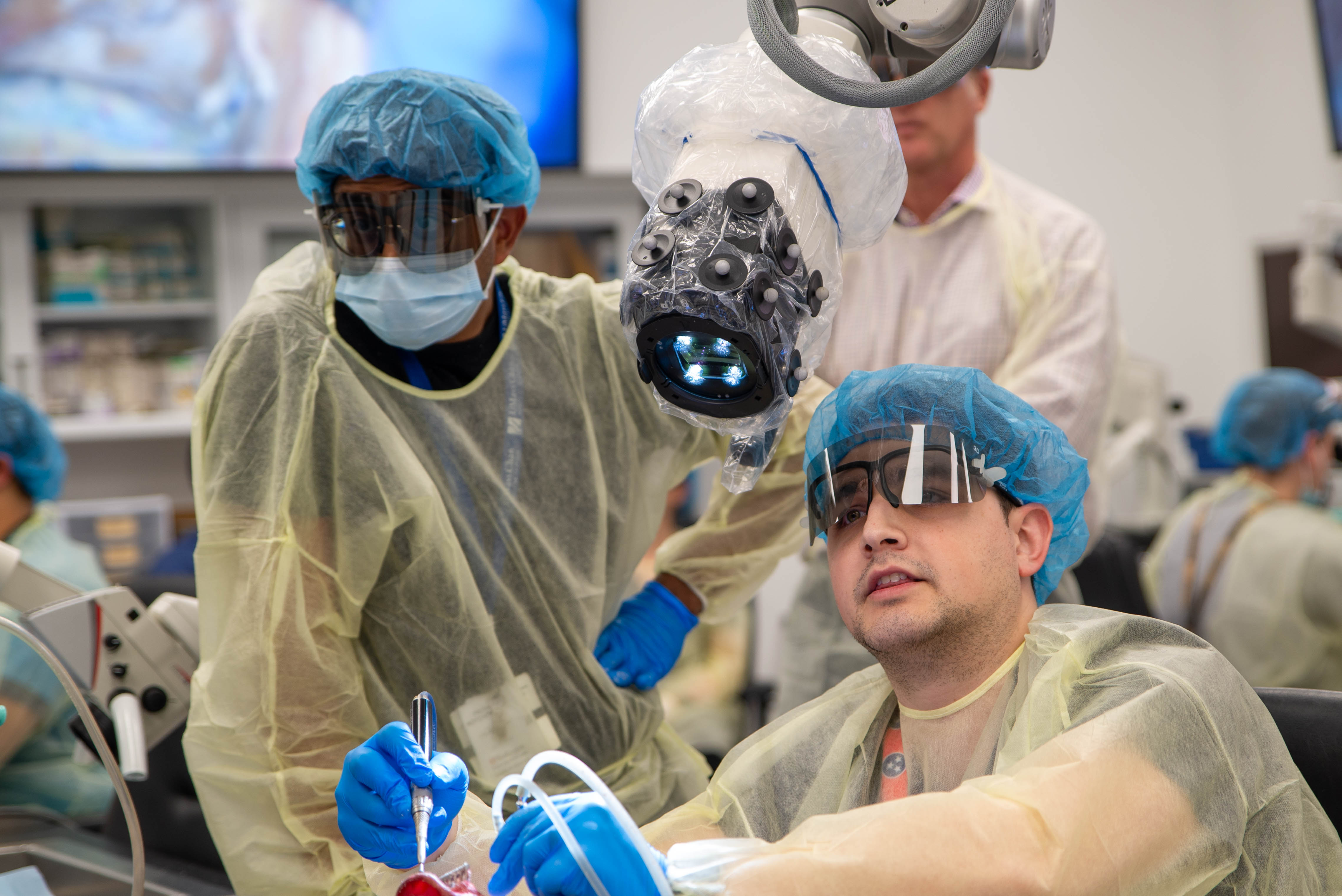
Additional vendor support offered residents experience with the latest technology in 3D surgical imaging, including the Synaptiv Modus X Exoscope and the Hoth Intel augmented reality platform.
Course co-director Matthew Shew, MD, was pleased with the collaborative experience the course provided.
“The joint neurosurgery-neurotology skull base course offers our trainees an invaluable, multidisciplinary learning experience that closely replicates the collaborative environment of the operating room,” said Shew. “This course not only allows participants to benefit from each other’s expertise but also highlights the importance of teamwork in complex surgical procedures. We were fortunate to have Dr. Madison, Dr. Chicoine, and Dr. Driscoll share their surgical expertise and, more importantly, the subtle nuances and lessons they’ve learned throughout their distinguished careers. At Washington University, our strong partnership with neurosurgery continues to elevate our research, education, and patient care.”